The Safest Implantation; The Third Stage of Dr. Yaman’s 3SE Methodology
A successful hair transplant is not limited to extracting grafts from the donor area. How the grafts are placed into the recipient area, how the hair follicles are protected throughout the procedure, and how a natural-looking result is created are also important. For this reason, Dr. Resul Yaman points to two main principles in the third stage of his 3SE methodology, The Safest Implantation. These two main principles are grafts survival rates and achieving a natural-looking result.
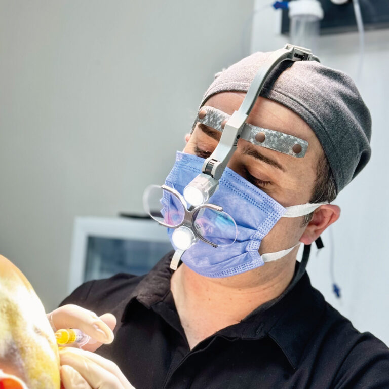
At Dr. Resul Yaman Hair Transplant Clinic, the DHI (Direct Hair Implantation) technique is applied during the implantation stage. Thanks to this method, the doctor is able to reduce the tissue trauma during graft placement, insert hair follicles at an appropriate depth, and create an individual implantation plan depending on anatomical features of the recipient area. However, every patient has different hair features, skin properties, hair loss pattern, and donor capacity. Safe implantation is not only related to the device used but also to the surgical plan of each individual case.
DHI Hair Transplant to Protect Grafts Survival Rates
In hair restoration, grafts survival rates are related to transplanted hair follicles continuing to grow after the healing process. During a hair restoration procedure, grafts are exposed to mechanical trauma when being extracted, stored, and placed in the recipient area. Mechanical trauma is one of the factors that affect the final result. That is why it is extremely important to choose such techniques that will protect graft integrity and viability as well as perform a careful surgical work.
According to Dr. Yaman, the grafts should be implanted in a controlled manner during the implantation stage. Therefore, he uses the DHI hair transplant technique in his clinic within the framework of his graft protection approach. In the DHI technique, the graft is placed inside the tube of the implanter device and implanted into the recipient area with a controlled motion. In contrast to the Classic FUE technique, the grafts are not held directly and planted using forceps.
In the 3SE The Safest Implantation methodology, the grafts are protected from mechanical effect. However, the survival rates of the grafts do not depend on the technique of their implantation alone. The method of extraction, the time spent out of the body, storage, surgical handling and the healing process of the patient also should be taken into account.
How Does the DHI Technique Protect Grafts and the Skin?
There are DHI implanters with different diameters. DHI implanter diameters vary from 0.7–1.1 mm and 0.9 mm devices are widely used. Due to the use of the small-diameter tools in hair implantation, it is possible to perform a minimally invasive procedure in the recipient tissue.
In the case of conventional forceps implantation, this diameter varies from 1.0–1.8 mm. For example, if implantation is conducted through 1 mm openings with the help of a DHI implanter pen for 4,000 grafts, the total width will be equal to 4 meters. When creating 1.5 mm openings for 4,000 grafts and performing forceps implantation, the scar will be 6 meters wide. That is why, the DHI technique provides a more skin-protective feature. However, these are only theoretical calculations aimed at explaining the total width of incisions.
In the DHI technique, the grafts are inserted through a tube rather than being held and pressed with forceps directly. Therefore, this method can be considered as a graft and tissue-protective implantation approach in patients who fit it.
Planning Angles, Direction, and Density for Natural-Looking Result
Another objective of safe implantation is to create a natural-looking hair. Not only proper graft placement in the right area is required to achieve it. Such factors as the exit angle of the hair, its direction, relationship with adjacent hair, and regional density should also be considered.
Dr. Resul Yaman pays special attention to controlling the angles and direction of the hair during implantation in the frontal hairline and temple areas. He uses the Yaman DHI Implanter in these regions. To perform a tissue-protective surgical procedure, the incisions corresponding to the implanter diameter are created with sapphire blades. It creates smoother incisions and protects the skin. Grafts are inserted using the Yaman DHI Implanter without the help of forceps.
Such an approach provides the possibility to plant the hair at lower angles and in the natural direction of its growth. It also helps to obtain a denser implantation according to the surgical plan. However, the density that can be achieved depends on the donor capacity of the patient and the recipient area characteristics.
Personalized Implantation Planning for Every Patient and Every Recipient Area
Every patient has different head shape, hair thickness, genetic hair loss pattern, and aesthetic expectations. That is why the application of the same implantation approach to every patient and every part of the recipient area is not appropriate.
Dr. Yaman takes not only individual patient characteristics but also the recipient area anatomical differences into account during the surgical planning. The frontal hairline, mid-scalp, and crown have different directions of the hair, angle requirements, and density target. That is why the implantation plan should be developed individually for each region.
Adjusting Graft Depth with DHI Implanters
To guarantee the safer graft implantation and better grafts survival rates, the grafts should be inserted at an appropriate depth. The depth can be adjusted both with the Yaman DHI Implanter and the standard DHI Implanter. Before the use of the Yaman DHI Implanter, the depth is adjusted with the help of sapphire blades. The standard DHI Implanter has a depth adjustment zone at the back of the device.
Dr. Yaman measures the length of the graft roots during the microscopic examination as well. As a result, the depth adjustment during implantation becomes easier.
Depth Control and the Microvascular Structure in the Crown Area
When placing the grafts into the crown area, the thickness of the scalp and tissue anatomy should be taken into account. The thickness of the skin in this area does not mean that the grafts should be inserted at a greater depth. One of the reasons why the crown procedures fail in some clinics is improper depth adjustment.
The hair follicles receive oxygen and nutrients via diffusion from the adjacent tissues. That is why the grafts should be placed at the depth that is favorable for this tissue environment. Although the crown area has thick skin, its dense microvascular structure is located close to the skin. Therefore, the insertion should be made right beneath the skin. If the grafts are inserted too deeply in the crown, they reach the larger blood vessels. Such vessels are not good for the diffusion-based nutrition.
Therefore, the sufficient insertion depth should reach the level of the smaller vessels supporting diffusion. On the other hand, the tissue structure of the frontal area is different. The dense microvascular structure is located a little deeper in the frontal area. For this reason, the graft implantation in the frontal area should be performed deeper than in the crown area.
Digital Mapping and CRM Follow-Up in Surgical Planning
The safe implantation begins from preoperative planning and continues after the surgery. The Dr. Resul Yaman Hair Transplant Clinic uses the CRM software for the surgical planning and patient follow-up.
During the surgery, the digital mapping of the donor and recipient areas is conducted using the clinical photographs. The donor area is divided into designated zones, and the planned amount of grafts to extract from each zone is recorded. In the similar way, the recipient area is divided into sections, and the planned amount of grafts for each section is recorded.
This information is displayed on the monitor in the operating room. Thus, Dr. Yaman and his surgical team can follow the planned graft distribution based on the same information. The monitoring of how many grafts will be inserted into each area helps the surgeon to perform the donor management in a controlled manner and stick to the surgical plan.
After the surgery, the patient is included in the CRM follow-up program. The assistant of Dr. Yaman requests the photographs at day 10 and at months 1, 3, 6, 9, and 12 after the surgery. The submitted photographs are analyzed to monitor the healing process, and the patient is informed about the findings requiring attention. This system helps to maintain communication after the surgery and monitor the hair transplant results during the first postoperative year.
Graft Distribution, Donor Management, and Personalized Hairline Design
A successful implantation plan requires using the available donor resources while considering long-term needs. The donor area is not an unlimited source of grafts. Therefore, the amount of grafts to allocate for the frontal region, mid-scalp, and crown areas should be carefully planned.
The hair thickness and multiple grafts rate are the important factors during the planning. The same number of grafts does not provide the same visual density in patients with fine and thick hair. The thick hair shafts give better visual density, while the fine hair cannot do this.
Dr. Resul Yaman develops the graft distribution according to the patient's donor capacity, hair features, and expectations of the final volume. The aim is to use the donor resources in the most efficient way, prioritize the areas that require this, and achieve a balanced and natural-looking result.
The hairline design also follows the personalized approach. The macro and micro irregularities are adjusted in order to create a natural hairline. The finer hairs are used in the transition zone, and the thicker grafts are placed behind them creating a gradual transition from the hairline to the greater density.
In suitable patients, the secondary beard grafts are mainly used for the implantation of the crown. This decision should be made taking into account the characteristics of grafts and the overall surgical plan.
Frequently Asked Questions About the Safest Implantation in DHI Hair Transplant
How does DHI hair transplantation help protect grafts?
The process involves the use of a special implanter to put the grafts in the recipient area. In this method, there is no need for using forceps to place grafts since it is done with an implanter. The safety of the grafts also depends on how they are extracted, stored, and handled.
Why is implantation depth important in hair transplantation?
Implantation depth affects how grafts interact with surrounding tissues. Placing the grafts too deeply or superficially can have negative implications in terms of healing and growing. Surgeons determine appropriate placement depth with graft characteristics, scalp thickness, and recipient-area anatomy.
How does Dr. Resul Yaman plan graft distribution?
Dr. Resul Yaman evaluates hair thickness, follicular unit composition, donor capacity, and recipient-area requirements. He plans graft distribution across the frontal region, mid-scalp, and crown to support balanced coverage while preserving donor resources for long-term needs.
What does the use of Yaman DHI implanter give in terms of natural result?
The surgeon uses the Yaman DHI implanter to help him for control graft placement, including angle and direction. The hair angles and directions are particularly important around the frontal hairline and temples. This gives a chance to achieve natural results, but it depends on individual characteristics and donor potential.
How does the implantation depth differ between the frontal region and crown?
The frontal region and crown have different anatomical characteristics and hair-growth patterns. When determining the implantation depth, the doctor and his team must take into account the thickness of the patient's scalp, the characteristics of the grafts, and the local tissue structure. A personalized approach will help to avoid wrong implantation depth.
How does the postoperative monitoring support hair transplant results?
The clinical team can assess patient's healing and hair growth over time with supporting the postoperative monitoring system. The patients need to send their photos at certain times during their first year to the clinic assistant. This helps the clinic stay in touch and see how their hair is growing.